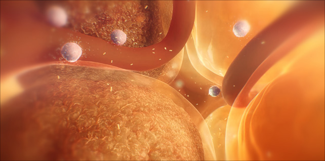
Fat cell enlargement & type 2 diabetes
Apr 22, 2022Fat cell enlargement in obesity is related to insulin resistance and type 2 diabetes.
In this sequence, we travel into the visceral fat and see an active environment – teeming with overstressed, damaged fat cells, vasculature, and immune cells.
Stress on these hypertrophic adipocytes results in lipid spillover (fatty acid release) into the circulation, as well as proinflammatory cytokine release and immune cell recruitment.
Tagged: adipocyte animation, adipocytes, adipose tissue disfunction, diabetes, diabetes 2, diabetes animation, diabetes prevention, fat cell animation, fat cell enlargement, fat cells, hybrid medical, hypertrophic adipocytes, hypertrophic adipose expansion, hypertrophic fat cells, medical animation, medical illustration, medical visualisation, scientific animation, type 2, type 2 diabetes
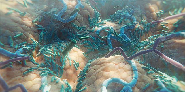
The diverse human gut microbiome
Jun 06, 2023Animation sequence featuring a fly-over of the human gut microbiome – a huge, diverse range of many different types of bacteria residing inside our small and large intestine.
This sequence was created for an episode (for WebMD) that examines how the gut microbiome may influence cancer response to immune checkpoint inhibitors. Essentially, if a patient has melanoma, the types of bacteria in their gut may help determine if the cancer responds to checkpoint inhibitor therapy or not.
There are trillions of microbes in our gastrointestinal tract, around 90% of which are bacteria.
Our microbiome has a profound impact on our well-being and can affect us in several different ways – both beneficial and harmful. In recent years, scientists and researchers have discovered correlations between the composition of an individual’s microbiome and their wider health, including obesity, mental health, bowel disease, and the immune system.
Further advances need to be made in learning more about actual causation – and which effects come from which individual microbes.
Tagged: bacteria, biology, biomedical communications, biotech, biotechnology, gastrointestinal tract, gut bacteria, gut diversity, gut health, gut microbes, gut microbiota, human gut microbiome, hybrid medical, medical animation, Medical science, medical visualization, medicine, microbiology, microbiome, microbiome health, microbiome medicine, pharma, science, science visuals, scientific animation, visual science
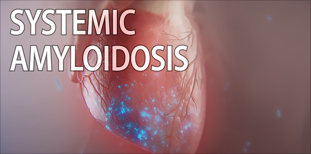
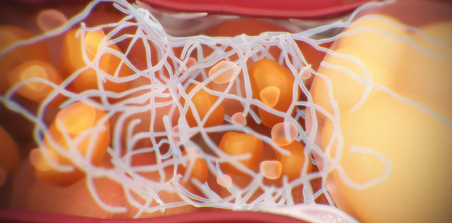
Systemic amyloidosis MOD
Jul 15, 2024A few excerpts from a 3-minute MOD animation that provides an insightful overview of amyloidosis, a group of rare and devastating progressive diseases caused by the accumulation of misfolded proteins.
The most common form of systemic amyloidosis is amyloid light chain (AL) amyloidosis, which is caused by the misfolding of immunoglobulin light chains produced by abnormal plasma cells in the bone marrow. These misfolded light chains aggregate, forming amyloid fibrils that accumulate in organs and tissues, leading to damage and dysfunction.
A crucial detail and task for us was to create an accurate depiction of the immunoglobulin light chain monomer, its conformational misfolding – and ultimately, its aggregation and assembly into amyloid fibrils – which accumulate into deposits and damage organs.
Given the significant impact on the heart (may lead to conditions such as cardiomyopathy in over 75% of patients), the focus of this animation is on illustrating the damage caused by amyloid deposits in cardiac tissue. While the heart is most commonly affected, amyloid fibrils can also accumulate in other organs such as the liver, wrists, hands, feet, tongue, and gastrointestinal tract.
By illustrating these key processes, this animation highlights the severe impact of amyloid deposits on the heart and other organs affected by AL amyloidosis.
Tagged: AL amyloidosis, amyloid deposits, amyloid light chain, amyloidosis, cardiac, cardiac amyloidosis, cardiology, fibrils, heart, heart failure, heart health, hybrid medical, immunoglobulin light chains, light chain amyloidosis, medical animation, medical marketing, misfolded proteins, misfolding, myocardium, scientific animation, visual science
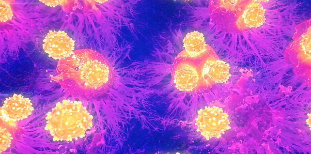
Cytotoxic T cells targeting tumor cells | immunotherapy animation
May 03, 2022A close-up fly-through of tumor cells, and the arrival of cytotoxic t cells intent on eradicating them. Activation and proliferation of cytotoxic T cells are critical for immune-mediated tumor destruction.
A key challenge we were given for this sequence: develop a rendering approach that mimics infrared thermal microscopy imaging techniques.
Researchers have been making advancements in understanding the thermal behavior of cells – for example, the ability to read the signatures of tumor cells due to their higher metabolism – in order to be used alongside existing histological analysis methods.
Tagged: biology, biomedical communications, biotechnology, cancer cells, cytotoxic T cells, fluorescence microscopy, hybrid medical, immune cells, immunology, immunotherapy, infrared, infrared (IR) microscopy, infrared imaging in biology, infrared spectroscopy, IR imaging, IR microscopy, Killer T cell animation, Killer T cells, medical animation, microspectroscopic animation, Minneapolis Minnesota, oncology, pharma, science visuals, scientific animation, T cells, thermal, thermal imaging, thermal microscopy, tumor cells, tumor death
TOTAL30® monthly replacement contact lenses
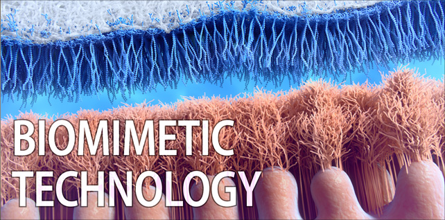
Feb 05, 2024Selected segments from a 2.5 minute animation that highlights the features and benefits of a new biomimetic technology that combines a biomimetic surface and unique lens chemistry to help resist bacteria and lipid deposits. Durable enough to deliver comfort for a full 30 days of wear, the TOTAL30 contact lens surface mimics the softness of the human cornea, keeping the eyes healthy with high oxygen permeability and superior lens surface moisture stability.
Unique challenges for this project included building a microworld of corneal epithelium and glycocalyx – a network of proteoglycans that covers epithelia of the eye – as well as construction of a polymer nanofiber “brush” that moves in a constant, dynamic, undulating motion like that of the corneal surface.
Another critical aspect was showcasing how these two dynamic surfaces (glycocalyx and polymer nanofibers) come together to form a layer of surface protection with the tear film. This biomimetic design provides a naturally clean, lubricious surface that is as soft as the human cornea, yet durable enough to last through 30 days of cleaning and maintenance.
Tagged: bacterial resistance, biomimetic technology, biomimicry, clean lens technology, contact lens animation, contact lenses, corneal care, corneal epithelium, digital health, dynamic surfaces, eye health, eye protection, glycocalyx, hybrid medical, lens chemistry, lens comfort, lens durability, lens innovation, long wear lenses, medical animation, medical device animation, medical marketing, moisture stability, oxygen permeability, pharma, polymer nanofibers, science communications, scientific animation, soft contact lenses, tear film protection, total 30 lens
Understanding CAR T-Cell Therapy: A Breakthrough in Cancer Treatment
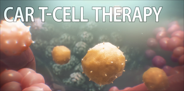
Nov 30, 2023This animation provides a straightforward yet comprehensive explanation of how CAR T-cell therapy represents a powerful new tool in the fight against cancer.
Our key task was to visualize a basic understanding of the immune system’s natural processes with these amazing scientific advancements that have led to the development of this innovative therapy.
CAR T-cell therapy is a cutting-edge approach to treating cancer by enhancing the body’s immune system. Normally, T-cells detect and destroy harmful invaders like bacteria, viruses, and cancer cells using specialized receptors. However, cancer cells can evade this process by causing T-cells to become exhausted or by blocking receptor binding, allowing tumors to grow unchecked.
The scientific advancements of this innovative therapy involve genetically modifying T-cells from a patient’s own body. Scientists insert DNA to create Chimeric Antigen Receptors (CARs), which are engineered to specifically recognize and bind to cancer cells. Once these modified T-cells – or CAR T-cells – are reintroduced into the patient’s bloodstream, they seek out and kill cancer cells more effectively. Additionally, these CAR T-cells do not just attack once. They multiply within the body, creating more fighters that can continue the assault against the cancer. This multiplication ensures a more robust and sustained attack, increasing the likelihood of successful treatment.
City of Hope is at the forefront of developing groundbreaking therapies that utilize CAR T-cells to treat various types of cancer, offering new hope to patients who might not have had effective treatment options before.
Tagged: (CAR) T-cell therapy, antigens, biology, biotechnology, cancer, cancer research, cancer treatment, CAR T, CAR T cell animation, CAR T therapy, CAR T-cell animation, car-t cell therapy, chimeric antigen receptor, city of hope, gene therapy, hybrid medical, immune health, immunotherapy, medical animation, Medical science, medical visualisation, pharma, scientific animation, T cells, visual science
Oligomers, fibrils, & plaques | Alzheimer’s | medical animation
Aug 21, 2023Segment from 5-minute mechanism of action animation.
This sequence explores a hallmark and pathophysiological feature of Alzheimer’s disease: toxic amyloid beta aggregates (Aβ) that accumulate into plaques (neurofibrillary tangles) near neurons in the brain.
A challenge for this sequence was to show the assembly of these naturally occurring peptides – oligomers, fibrils, and plaques – all in one continuous shot.
In addition, we show how over time, extracellular amyloid beta aggregates, including oligomers, fibrils, and plaques, are thought to exert toxic effects on neurons and synapses, disrupt cell function, and lead to synaptic dysfunction and loss.
Tagged: aggregates, alzheimer’s, alzheimer's disease, alzheimers, amyloid, amyloid beta, amyloid beta plaques, amyloid fibril, amyloid plaques, biomedical communications, biotech, brain deterioration, cognitive decline, digital health, extracellular amyloid beta aggregates, fibrils, hybrid medical, medart, medical animation, medical visualization, neurofibrillary tangles, neuroscience, oligomers, plaques, scicomm, synaptic dysfunction, toxic aggregates
Amyloid light chain (AL) amyloidosis | cardiac amyloidosis animation
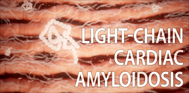
Jul 21, 2023Opening with a view of the heart, we reveal several amyloid deposits infiltrating the muscle tissue.
Then we zoom down to the heart muscle cells and see misfolded light chains aggregating into insoluble amyloid fibrils.
We fade, over time, from the early onset of light-chain infiltration to late-stage fibril aggregation, where the buildup of amyloid deposits around and in between the cardiomyocytes has made the muscle tissue stiff and less flexible, impairing the heart’s ability to relax and fill with blood.
Amyloid light chain (AL) amyloidosis is the most common form of systemic amyloidosis and is caused by the misfolding of immunoglobulin light chains, which are produced by abnormal plasma cells in the bone marrow.
Deposits can be widespread, affecting multiple organs, or they can be more localized.
Tagged: AL amyloidosis, amyloid deposits, amyloid light chain, amyloidosis, cardiac, cardiac amyloidosis, cardiology, fibrils, heart, heart failure, heart health, hybrid medical, immunoglobulin light chains, light chain amyloidosis, medical animation, misfolded proteins, misfolding, myocardium, scientific animation, visual science
Non-alcoholic steatohepatitis (NASH) | NASH liver disease animation
May 16, 2022A few selected sequences we produced for a recent Medscape CME / ABIM MOC / CE symposium titled NASH 101: What You Need to Know Now
The full 2.5- minute animation was developed to be played as a visual aid when the moderator discussed epidemiology and pathogenesis.
We were asked to conceptualize and develop this visual science story. It provides insight into the pathophysiology of NAFLD and NASH, including the role of fatty acids, insulin resistance, and steatosis, as well as mitochondrial dysfunction, excess ROS production, and the stress put on the endoplasmic reticulum inside ballooning hepatocytes.
In addition, we depicted key attributes of the progression of non-alcoholic steatohepatitis (NASH) including bloated hepatocytes and the fat droplets accumulating inside, displaced cell organelles, lobular inflammation, and the scarring (fibrosis) that occurs as collagen fibers fill the spaces once cell death occurs.
In the last few years, nonalcoholic fatty liver disease (NAFLD) and nonalcoholic steatohepatitis (NASH) have become the most common causes of chronic liver disease worldwide. This epidemic is starting to parallel the epidemics of obesity and type 2 diabetes.
Tagged: biology, biomedical communications, cirrhosis, fatty acids, fatty liver disease animation, hepatocytes, hybrid medical, insulin resistance, lipotoxicity, liver animation, medical animation, medical marketing, NAFLD, NASH, NASH liver disease animation, non-alcoholic steatohepatitis, nonalcoholic fatty liver disease, obesity, science visuals, scientific animation, steatohepatitis, steatosis, type 2 diabetes
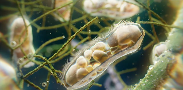
Paramecia: unicellular ciliates animation
Jun 02, 2023A view of paramecia spiraling their way about in a freshwater pond.
In this sequence, we get a close-up look at one of these unicellular microorganisms as it makes its way around using the tiny hair-like cilia on its outer surface. Arranged in longitudinal rows, there are around 4,000 of these motile cilia on its surface, beating in waves to ensure movement and feeding.
In addition to other organelles, we can see several vacuoles inside, digesting organisms such as bacteria and yeasts.
The paramecium was among the first single-celled organisms to be observed under the microscope.
In order to capture the emergent behavior and movement of this microworld, the definitive goal here was to create a natural environment using physics simulations and a procedural pipeline instead of hand-keyed animation.
Tagged: biology, cell, ciliate, ciliates, houdini, houdinifx, hybrid medical, medical animation, micro, microbes, microbiology, microcosmos, microscope, microscope photography, microscopy, microworld, paramecia, paramecium, pharma, photomicrography, redshift, redshift3d, science, science visuals, scientific animation, sidefx, unicellular
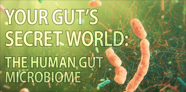
Your Gut’s Secret World: The Gut Microbiome
Nov 19, 2024The relationship between your gut microbiome and your well-being is so extensive that almost no condition is untouched by its influence. What is the gut microbiome, and how does it impact your overall health?
Join us as we explore the uniquely individual ecosystem that is your gut microbiome, and the intricate environment within the human gut, which supports essential functions such as digestion, vitamin synthesis, pathogen defense, immune response, and even mental health.
Our journey begins within the inner mucus layer of the descending colon, where we observe essential bacterial byproducts — including short-chain fatty acids, hormones, and dietary nutrients — being absorbed by the colon’s epithelial cells. Immune cells are also here, patrolling the area and maintaining a delicate balance of tolerance versus response within the gut ecosystem. This inner mucus layer serves as a primary defense barrier, limiting direct microbial contact with the colon.
Next, we transition to the permeable outer mucus layer, a vast, thriving habitat for microbial colonization and interaction. Of the 100 trillion microbes coexisting in your gut, the colon hosts the most robust and diverse population.. Fungi and viruses – especially bacteriophages – also play significant roles in this dynamic environment.
Finally, we explore a defining feature of the microbiome: biofilms. These structured colonies of bacteria produce protective coverings and attach to the inner mucus layer. Biofilms play a fundamental role in microbial life, contributing to both health and disease. Understanding the balance between beneficial and harmful biofilms is crucial for developing strategies to manage gut health effectively.
Tagged: bacteria, biomedical communications, biotech, biotechnology, colon health, gastrointestinal tract, gut bacteria, gut diversity, gut ecosystem, gut health, gut microbes, gut microbiome, gut microbiota, human gut microbiome, human microbiome, hybrid medical, medical animation, Medical science, medical visualization, medicine, microbiology, microbiome, microbiome health, microbiome medicine, microbiota, pharma, science, science visuals, scientific animation, visual science
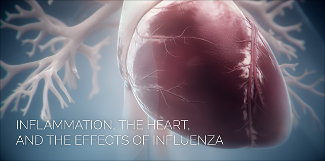
Inflammation, the Heart, and the Effects of Influenza
Mar 15, 2022Sequence from an animation that explores influenza, the body’s immune response to the virus, and how it may cause dangerous levels of inflammation throughout multiple organs systems of the body.
In this excerpt, we depict a plaque rupture and show the important role inflammation plays in the development of atherosclerotic plaques.
This is a sequence from a three-part production that provides insight into how older adults with existing conditions such as cardiovascular disease, respiratory illness, or diabetes may face a higher risk of severe complications when they are infected with the influenza virus.
Tagged: atherosclerosis, cardiac, cardiac arrest, cardiology, cardiovascular disease, flu, heart disease, heart disease awareness, heart failure, hybrid medical, hypertension, immune response, inflammation, influenza, medical animation, plaque, scientific animation, vaccines, virus, visual science

Onychomycosis & nail fungal disease
May 22, 2022This animation takes a closer look at a fungal infection of the toenail and the key attributes of its invasion, including discoloration and brittleness of the nail, striations and thickness of the nail plate, and ultimately, the presence and proliferation of fungi at the nail bed.
One of the more important aspects for this project was to create a highly detailed, diseased nail cross-section, including anatomically correct layers (dorsal, intermediate, and ventral). This would allow us to feature the delivery vehicle and transport of cosmetic and antifungal ingredients through the nail plate.
Additional key challenges for this piece included creating a 3D toe with diseased nail, particle simulations of the key anti-fungal solution ingredients and their pathway down to the nail bed, and the destruction of fungal species (including yeasts, dermatophyte molds, and non-dermatophyte molds) at the nail bed.
Trichophyton rubrum – the fungal species we see at the end – is the most common type of nail fungus.
Tagged: antifungal, cosmetic, dermatology, dermatophyte, fungal, fungal infection, fungal nails, fungus, houdini, hybrid medical, keratin matrix, medical animation, mold, nail bed, nail fungal disease, nail plate, nails, onychomycosis, pedicure, redshift, science, science visuals, scientific animation, toe fungus, toe fungus animation, toe fungus3D, toenail fungus, trichophyton, trichophyton rubrum, yeast
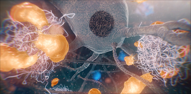
Proliferation and activation of microglia in the brain
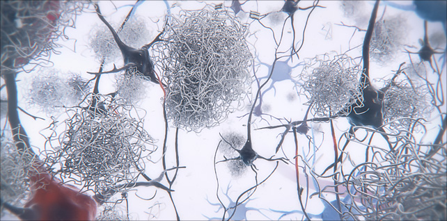
Aug 31, 2023Amyloid plaques are the hallmark of Alzheimer’s disease (AD).
This sequence shows the proliferation and activation of microglia in the brain, concentrating around and engulfing neurotoxic amyloid-beta plaques that have caused damage to impaired neurons nearby.
Microglia are specialized immune cells of the central nervous system, specifically in the brain and spinal cord. They play a vital role in monitoring and maintaining the neural environment. One of their primary responsibilities is to detect and remove damaged neurons, plaques, and other cellular debris from the brain.
We worked closely with our clients to create an accurate portrayal of microglia, including their form and structure, their movement and behavior, as well as the manner in which they form a barrier around these growing, toxic plaques. Microglia size, movement, and morphology were all examined by viewing time-lapse imaging projection software – and then recreated in 3D.
One potentially harmful aspect of this relationship between microglia and plaques is that prolonged activation of microglia in response to persistent plaques can lead to chronic inflammation, which might contribute to neuronal damage and the progression of Alzheimer’s disease.
Tagged: aggregates, Alzheimer’s disease, alzheimers, amyloid, amyloid beta, amyloid fibril, amyloid plaques, amyloid-beta plaques, axonal dystrophy, Aβ plaques in Alzheimer’s disease, biomedical communications, biotech, biotechnology, brain deterioration, cognitive decline, glia, hybrid medical, medical animation, medical visualization, microglia, microglia barrier, microglial, microglial phagocytosis, Minneapolis Minnesota, nervous system, neurofibrillary tangles, neurons, neuroprotection, neurotoxic, plaques, scientific animation, synaptic dysfunction, toxic aggregates